


As the most obvious symptoms that show diabetic microangiopathy, diabetic retinopathy (DR) is featured by a fundus disease with specific changes as well as one of the serious complications of diabetes.
Based on the sign that whether there is retinal neovascularization clinically or not, diabetic retinopathy can be divided into two parts: non-proliferative diabetic retinopathy (NPDR), diabetic retinopathy without retinal neovascularization, and proliferative diabetic retinopathy (PDR).
Causes
Due to the insulin metabolism disorders that lead to changes in ocular tissues, nerves and blood vessels, people with diabetes are prone to suffer from damages to ocular nutrition and visual function. Microvascular is between the small arteries and veins minor while lumen is less than 100-150µm in fine capillary network and vascular.
Besides, it provides room for tissue and blood to change materials. Changes in blood composition enable the vascular endothelial cells to function abnormally, hereby resulting in the blood-retinal barrier.
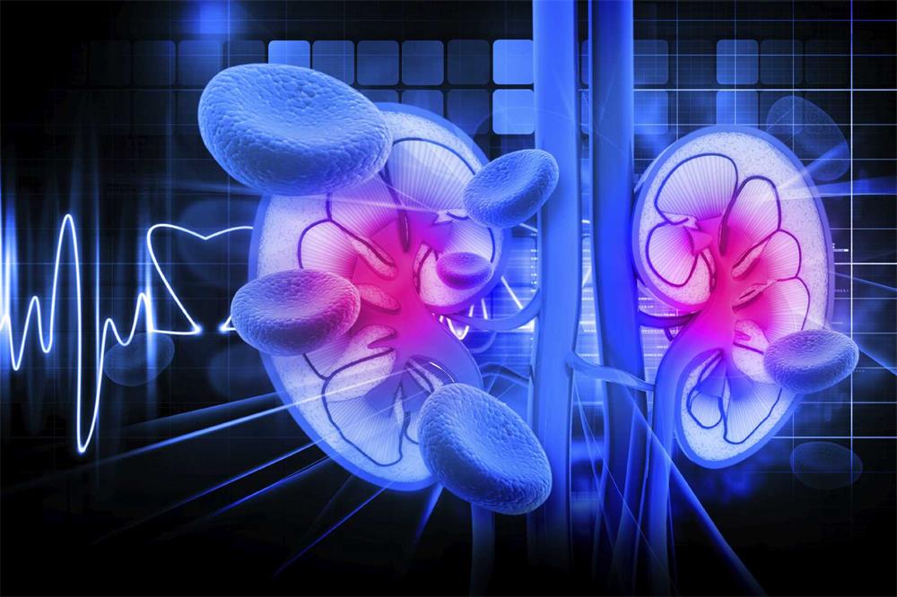
The destroy of the pigment epithelial cells of the retinal capillary endothelial cells causes the leakage of small blood vessels. Symptoms can be founded in the part of retina and kidney. However, this is the main reason of blindness, renal failure and even death.
Symptoms
The symptoms of retinal capillaries cover aneurysms, bleeding spots, hard exudates, cotton wool spots, beaded veins, intraretinal microvascular abnormalities (IRMA), and macular edema. Extensive ischemia can lead to retinal and optic disc neovascularization, preretinal hemorrhage and traction retinal detachment. In addition, the patient has a severe visual impairment.
It's required to check whether there is diabetic nephropathy in time.
3. Cholesterol and blood lipid check
There is a need to detect cholesterol and blood lipid levels.
4. Fundus fluorescein angiography check
If diabetic retinopathy has not been found under the ophthalmoscope, the fundus fluorescein angiography may show abnormal fluorescence patterns. The microvascular tumors under fundus fluorescein angiography are more numerous than those found under ophthalmoscopy.
5. Electroretinogram oscillatory potentials (OPs)
As a subcomponent of electroretinogram (ERG), OPs can objectively and boast sensitivity to reflect the blood circulation status of the inner retina. According to the eyes with no disease on the fundus, it depicts the abnormal amplitude of OPs. While people with diabetic retinopathy, it can further monitor the process and improvement of diseases.
6. Other checks
Visual contrast sensitivity check, blood viscosity check and serum SOD activity check are also helpers.
Treatments
1. Medical treatment
2. Photocoagulation treatment
Laser treatment is thought of as an effective method for treating diabetic retinopathy. It has been proven that photocoagulation therapy delivers beneficial results to the pathogenesis of the the disease. On the one hand, it can enable the degeneration and regeneration of new blood vessels. On the other hand, it can reduce edema.
3. Cold treatment
Condensation is mainly designed for people who are not suitable for photocoagulation therapy and supplementary therapy for photocoagulation therapy. Patients suffer from refractive interstitial turbidity and peripheral retinal lesions that cannot be treated by photocoagulation.
4. Vitrectomy
The basic indications for vitrectomy are vitreous hemorrhage and severe proliferative disease. It is generally believed that vitrectomy is required for patients with extensive vitreous hemorrhage for more than 3 months and failure to be absorbed on their own.
Conclusions
Causes, symptoms, checks and treatments are expounded in this article, which gives people a helper to know diabetic retinopathy in a comprehensive manner. It is recommended to see a doctor when the happening of DR.
Based on the sign that whether there is retinal neovascularization clinically or not, diabetic retinopathy can be divided into two parts: non-proliferative diabetic retinopathy (NPDR), diabetic retinopathy without retinal neovascularization, and proliferative diabetic retinopathy (PDR).
Causes
Due to the insulin metabolism disorders that lead to changes in ocular tissues, nerves and blood vessels, people with diabetes are prone to suffer from damages to ocular nutrition and visual function. Microvascular is between the small arteries and veins minor while lumen is less than 100-150µm in fine capillary network and vascular.
Besides, it provides room for tissue and blood to change materials. Changes in blood composition enable the vascular endothelial cells to function abnormally, hereby resulting in the blood-retinal barrier.
The destroy of the pigment epithelial cells of the retinal capillary endothelial cells causes the leakage of small blood vessels. Symptoms can be founded in the part of retina and kidney. However, this is the main reason of blindness, renal failure and even death.
Symptoms
The symptoms of retinal capillaries cover aneurysms, bleeding spots, hard exudates, cotton wool spots, beaded veins, intraretinal microvascular abnormalities (IRMA), and macular edema. Extensive ischemia can lead to retinal and optic disc neovascularization, preretinal hemorrhage and traction retinal detachment. In addition, the patient has a severe visual impairment.

PDR causes retinal damage and then stimulates the growth of new blood vessels, which is harmful to the retina. Fibrosis and even retinal detachment can be brought. In the meanwhile, new blood vessels grow into the vitreous, leading the hemorrhage to happen. Compared with NPDR, PDR can threaten vision loss and even complete blindness.
Checks
1. Blood sugar check
Regularly measuring blood sugar levels is a must to monitor the development of diabetes.
It's required to check whether there is diabetic nephropathy in time.
3. Cholesterol and blood lipid check
There is a need to detect cholesterol and blood lipid levels.
4. Fundus fluorescein angiography check
If diabetic retinopathy has not been found under the ophthalmoscope, the fundus fluorescein angiography may show abnormal fluorescence patterns. The microvascular tumors under fundus fluorescein angiography are more numerous than those found under ophthalmoscopy.
5. Electroretinogram oscillatory potentials (OPs)
As a subcomponent of electroretinogram (ERG), OPs can objectively and boast sensitivity to reflect the blood circulation status of the inner retina. According to the eyes with no disease on the fundus, it depicts the abnormal amplitude of OPs. While people with diabetic retinopathy, it can further monitor the process and improvement of diseases.
6. Other checks
Visual contrast sensitivity check, blood viscosity check and serum SOD activity check are also helpers.
Treatments
1. Medical treatment
- To better manage diabetes
The treatment of diabetes is the root cause to treat diabetic retinopathy. Blood glucose control should be put in the first place in principle.

- To lower blood lipid
- To control blood pressure
2. Photocoagulation treatment
Laser treatment is thought of as an effective method for treating diabetic retinopathy. It has been proven that photocoagulation therapy delivers beneficial results to the pathogenesis of the the disease. On the one hand, it can enable the degeneration and regeneration of new blood vessels. On the other hand, it can reduce edema.
3. Cold treatment
Condensation is mainly designed for people who are not suitable for photocoagulation therapy and supplementary therapy for photocoagulation therapy. Patients suffer from refractive interstitial turbidity and peripheral retinal lesions that cannot be treated by photocoagulation.
4. Vitrectomy
The basic indications for vitrectomy are vitreous hemorrhage and severe proliferative disease. It is generally believed that vitrectomy is required for patients with extensive vitreous hemorrhage for more than 3 months and failure to be absorbed on their own.
Conclusions
Causes, symptoms, checks and treatments are expounded in this article, which gives people a helper to know diabetic retinopathy in a comprehensive manner. It is recommended to see a doctor when the happening of DR.
Leave a comment
All comments are moderated before being published.
This site is protected by hCaptcha and the hCaptcha Privacy Policy and Terms of Service apply.